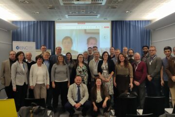
Phase transitions

High-temperature neutron diffraction study of the phase transition in a system of lead and bismuth-based perovskites

measured by the PSD and MTEST

Neutron powder diffraction (NPD) was performed to determine the crystalline and magnetic structure of Pb0.5La0.5Fe1-xMnxO3 (0≤x≤1.0) and Pb0.5-xBixLa0.5FeO3 (0<x≤0.25) transition metal oxide system. Due to difficulties in high-grade sample preparation the structure of this system had been poorly resolved. PbyLa1-yMnO3 (y≤0.5) is a ferromagnets with Curie temperature TC increasing linearly with x to 355K for x=0.5 and exhibiting colossal magnetoresistance (CMR) with  an MR value of 50% in a magnetic field of 5.5 T at 330 K in thin films. Doping the Mn sites with Fe dramatically alters the material properties leading to variety of applications of Pb1-yLayFe1-xMnxO3 as catalysts, electrode materials in solid oxide fuel cells, exhaust gas sensors and membranes for separation processes etc.

Results

The powder samples with compositions (according to ICP analyses) close to the nominal formula were successfully synthetized by a specific solution combustion technique and characterized by thermo-gravimetric/differential thermal analysis (TG/DTA), X-ray diffraction (XRD), magnetometry and Mössbauer spectroscopy (MS).

The neutron diffractometers PSD (λ0=1.061 Ǻ) and MTEST (λ=1.439 Ǻ) were used. The allotted beam time was only sufficient to take NPD patterns at 295 K and elevated temperatures on the system Pb0.5La0.5Fe1-xMnxO3 (0≤x≤1.0). The XRD analyses showed that the samples were single phases with apparently pseudo cubic perovskite structure. The XRD patterns could be indexed in the R-3c rhombohedral space group but structure refinements using FullProf produced high χ2.  Susceptibility, TG/DTA and NPD data (Fig.1) evidenced that the Néel temperature (TN≈555 K, x=0) goes down with increasing x.

Fig 1

Figure 1. Neutron powder diffraction pattern for Pb0.5La0.5Fe1-xMnxO3 (0≤x≤1.0) and for Pb0.25Bi0.25La0.5FeO3

Simultaneous XRD and NPD Rietveld analyses showed that La0.5Pb0.5FeO3 (x=0) is best described in orthorhombic Pnma space group (Fig. 2) in similarity with LaFeO3.

Fig 2

        Figure 2. Neutron diffraction pattern and Rietveld refinement for La0.5Pb0.5FeO3

The rest crystal structures had Imma symmetry (except R-3c for x=1) as exemplified in Fig. 3 for x=0.75. Though Mn3+ and Fe3+ are with practically identical ionic radius (0.645 Ǻ), the doping affects the crystal structure: there are sizeable lattice deformations reflected in changes in unit cell volume, bond angles and cation-oxygen distances. Furthermore, the magnetic interactions are strongly influenced: the Mn rich compounds (x > 0.5) display ferromagnetism whereas those with high Fe content are canted antiferromagnets. The magnetic structure of x=0 is of GxFz-type with main component of the effective magnetic moment on the iron site Sx = 3.6± 0.2 μB.

Fig 3

    Figure 3. Neutron diffraction pattern and Rietveld refinement for Pb0.5La0.5Fe0.25Mn0.75O3